Human chronological or biological age doesn’t directly show the real body maturation stage, hereditary, functional, environmental, nutritional, sexual, metabolic, social, emotional, cultural factors, affect the development.
Table of Contents
Introduction
Human chronological or biological age doesn’t directly show the real body maturation stage, hereditary, functional, environmental, nutritional, sexual, metabolic, social, emotional, cultural factors, affect the development. Third molar radiographic examination which determines it's presence, position and degree of calcification, morphology and eruption, effect on the dental arch development is commonly used in oral surgery, orthodontics and pediatrics for diagnosis and treatment planning.
Intra-Extra Oral Radiographs required
- Accurate Periapical Film
This help in evaluating the position of impacted tooth in relation to the adjacent ones, and also show shape and size of roots, the tooth relationship to adjacent anatomical structures e.g Inferior Alveolar Canal .
- Occlusal Film
Only supplementary to periapical film. it reveals Bucco-Lingual position of at least the crown of the impacted tooth.
- Bitewing Radiograph
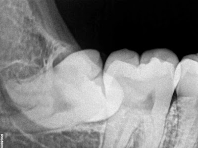
It has been used in Class I&II cases of impacted 3rd molars. It has acurate visualization of relationship between the crown of 3rd molar and the 2nd molar as it is directed with right angle x-ray "0 degree".
- Orthopantomograph "OPG"
This is a very helpful study which will sho the impacted tooth with its relations to the adjacent teeth as well as the anatomical land- marks, shape and the amount of covering the impactions as well as the shape and size of the jaw bone.
- Lateral Radiographic View of mandible
A more accurate roentgenogram in class II horizontal impacted third molar is obtained by a correctly positioned lateral view of the mandible not the oblique view.
- Tube shift method
Helps in localization the mandibular canal in relation to the apices of the lower third molar. Frank suggested that a modification of the A tube-shif" method can be used to deter mine whether the mandibular canal is medial to, lateral to or below an impacted mandibu- lar third molar. This technique was first described by Richards. The principle involved is the same as that of the "clark's shift" to localize a maxillary impacted cuspid.
